
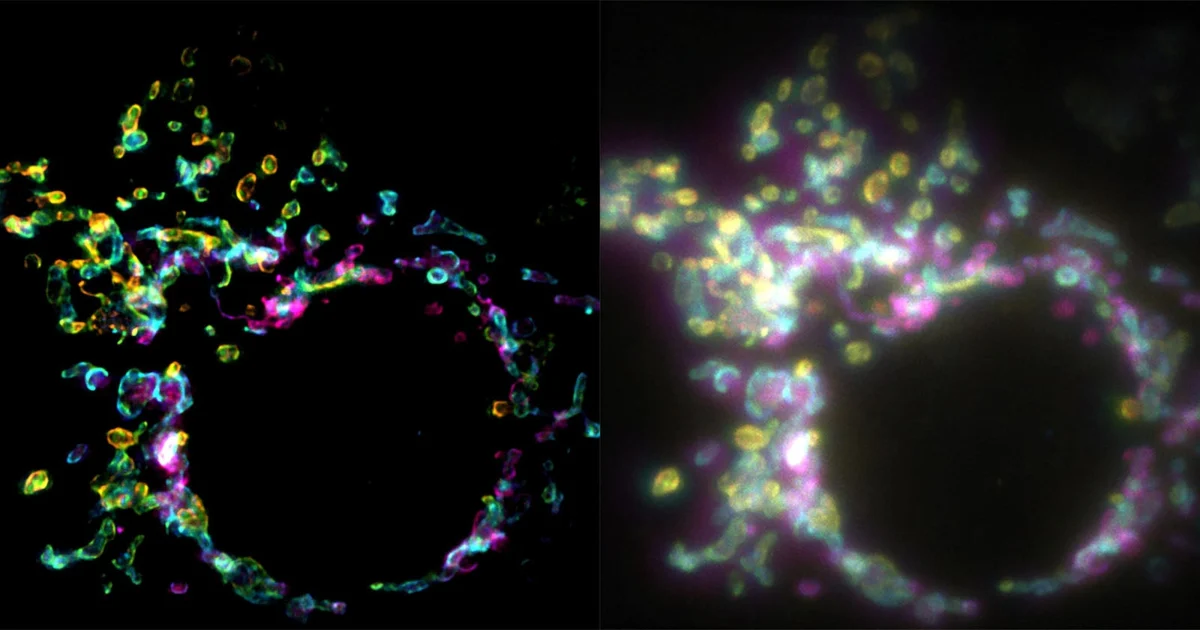
A groundbreaking advancement in optical microscopy has emerged from the Laboratory of Nanoscale Biology (LBEN) at the École Polytechnique Fédérale de Lausanne (EPFL), promising to redefine how researchers visualize the dynamic, often chaotic environment inside living cells. The new technique, dubbed SPIFFI—or spatial polarization-induced fluorescence fluctuation imaging—allows for the generation of super-resolution images from a single exposure, effectively circumventing the limitations that have historically hindered the study of fast-moving biological processes.
For decades, the field of cell biology has struggled to bridge the gap between high-resolution detail and temporal speed. While traditional light microscopy provides a window into the cell, it is governed by the Abbe diffraction limit, which restricts resolution to approximately 200–300 nanometers. Super-resolution techniques developed over the last twenty years have successfully broken this barrier, yet they have typically come at the cost of speed. SPIFFI, as detailed in the recent publication in the journal Nature, represents a fundamental shift in this paradigm.
The Limitation of Temporal Resolution
To understand the significance of SPIFFI, one must look at the evolution of super-resolution microscopy. Methods such as Photoactivated Localization Microscopy (PALM) and Stochastic Optical Reconstruction Microscopy (STORM) have been revolutionary, earning their developers the Nobel Prize in Chemistry in 2014. These techniques rely on the sequential imaging of thousands of individual fluorophores, which are then computationally stitched together to form a single, high-resolution static image.
The inherent flaw in these existing methods is their reliance on "temporal accumulation." Because these techniques require the capture of thousands of frames to reconstruct a single clear image, they are effectively blind to the rapid, flickering movements occurring within living tissue. By the time a high-resolution image is reconstructed, the cell has already moved, warped, or undergone structural changes, rendering the resulting data a composite of blurred, outdated information. For scientists studying processes like mitochondrial fission, protein trafficking, or the rapid rearrangement of the cytoskeleton, current super-resolution techniques are often too slow to provide a meaningful look at these "live-action" events.
How SPIFFI Changes the Imaging Paradigm
The SPIFFI technique, spearheaded by researchers Wei Guo, Lely Feletti, and Professor Aleksandra Radenovic, approaches the problem through a different physical property of light: polarization. Fluorescent molecules, or fluorophores, do not emit light uniformly in all directions; rather, they oscillate in specific planes based on their orientation within the cellular architecture.
SPIFFI exploits this by utilizing a specialized optical setup that splits fluorescent light into four distinct, polarization-sensitive channels. By comparing these channels simultaneously, the system can derive structural information that is otherwise buried in the background noise of conventional widefield images. Because this data is captured in a single frame, the requirement to record hundreds of images to "build" the resolution is eliminated.
According to the research team, this methodology improves spatial resolution by a factor of two in a single pass. While standard diffraction-limited images might show a vague, glowing blob, a SPIFFI-processed image reveals the crisp, defined edges of cellular structures at a resolution of approximately 160–170 nanometers. When further integrated with existing fluctuation-based post-processing algorithms, the team has demonstrated the ability to reach resolutions as fine as 80 nanometers, all while maintaining a frame rate sufficient to record video.
Chronology of Development and Experimental Success
The development of SPIFFI represents the culmination of years of work within the LBEN at EPFL. The team began by identifying that the primary bottleneck in super-resolution was not the lack of light, but the lack of information density in the time domain.
In early 2026, the team focused on optimizing the polarization-splitting hardware. By mid-2026, they had successfully tested the technique on mitochondrial outer membranes, a notoriously difficult structure to image due to their constant movement and fusion. The results were stark: where traditional widefield microscopy showed a smeared, blurred representation of the mitochondria, SPIFFI produced high-contrast, sharp images that allowed the researchers to observe the precise moments of mitochondrial fission and fusion.
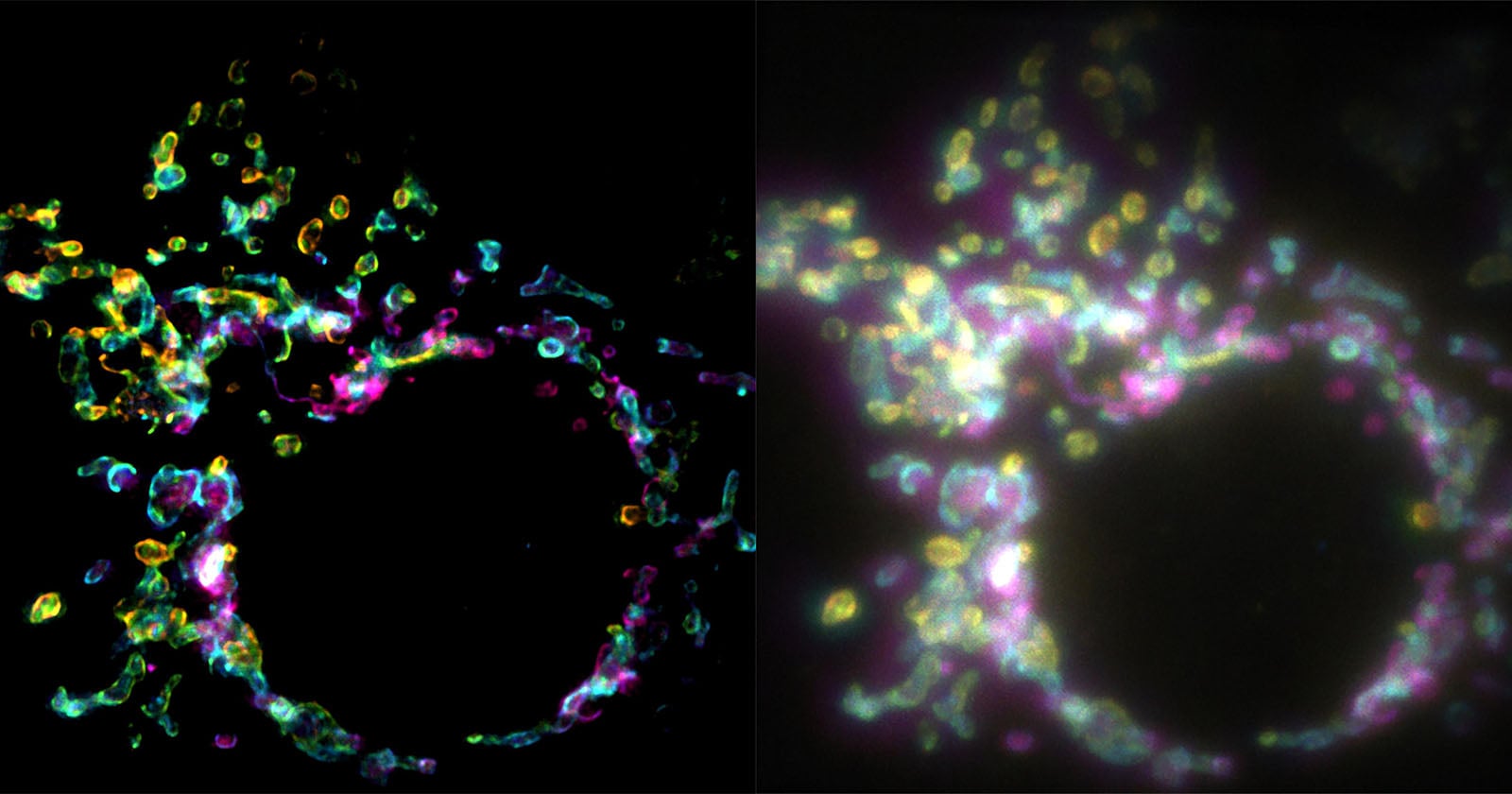
This success confirmed that SPIFFI could maintain the "super-resolution" tag while operating at the speeds necessary for live-cell imaging. The subsequent publication in Nature serves as the formal validation of this methodology, providing the scientific community with the protocols to implement the technique.
Official Perspectives and Academic Response
"Essentially, previous approaches used temporal information to resolve spatial resolution, but this doesn’t work very well on living cells," notes Wei Guo, the lead author of the study and a PhD student at LBEN. His assessment highlights the central frustration of modern microscopy: the trade-off between clarity and time.
Professor Aleksandra Radenovic, who oversaw the project, emphasizes the practicality of the new method. "SPIFFI can capture fast-moving processes within cells, while enabling high-throughput, multi-dimensional imaging beyond the limits of conventional microscopes," she explained in a statement following the publication.
The academic community has received the findings with cautious optimism. Imaging specialists suggest that the most compelling aspect of SPIFFI is not necessarily the resolution limit itself—which is already approached by other methods—but the ease of integration. Many high-end microscopy setups in research institutions worldwide are currently limited by their inability to handle high-speed, high-resolution tasks. SPIFFI’s hardware—which involves a modular optical splitter—is designed to be retrofitted onto existing fluorescence microscopes. This makes it an economically viable path to upgrading existing lab infrastructure rather than requiring the purchase of multi-million dollar, purpose-built systems.
Broader Implications for Biological Research
The implications of this technology extend far beyond the laboratory in Lausanne. If SPIFFI becomes a standard tool in biology labs, it will allow for a deeper understanding of cellular pathology. Many diseases, ranging from neurodegenerative disorders to cancer, are driven by the failure of cellular components to function or move correctly.
For instance, in the study of neurodegeneration, researchers often look for "traffic jams" of organelles within neurons. If these events are too fast to be captured by standard super-resolution techniques, they remain invisible. With SPIFFI, these transient, disease-linked structural shifts could be recorded, analyzed, and modeled with unprecedented clarity.
Furthermore, the technique offers a new frontier for pharmaceutical research. When testing the efficacy of a new drug, scientists often need to see how a compound affects the sub-cellular structure of a cell in real-time. The ability to watch a drug "in action" at a resolution of 80 nanometers, rather than waiting for fixed-cell snapshots, could significantly accelerate the development of targeted therapies.
Future Outlook and Technical Challenges
Despite the success of the initial experiments, the research team at EPFL is not resting on these results. Current efforts are focused on miniaturization. The existing optical hardware, while effective, still adds a layer of complexity to the microscope’s light path. To ensure widespread adoption, the team is working to create a more compact, plug-and-play version of the SPIFFI hardware.
Additionally, the computational side of the project—the algorithms that deconvolve the polarization data—is expected to see further refinements. As machine learning models become more adept at image processing, it is highly likely that the post-processing speed and resolution limits of SPIFFI will continue to improve.
As the technology moves from the research phase to potential commercialization, the microscopy market may see a shift. Laboratories that have been priced out of the super-resolution market due to the high cost and maintenance requirements of traditional systems may find in SPIFFI an accessible solution. By democratizing access to high-speed, high-resolution imaging, the work of Guo, Feletti, and Radenovic could potentially trigger a wave of new discoveries across the biological sciences, finally giving scientists a clear look at the intricate, fast-paced world that sustains life.