3d eye scans can detect signs of parkinsons – 3D eye scans can detect signs of Parkinson’s, a revolutionary development in the fight against this debilitating disease. While traditional methods often rely on symptoms that appear in later stages, these scans offer a potential window into the early stages of Parkinson’s, allowing for earlier diagnosis and treatment.
The technology utilizes advanced imaging to analyze subtle changes in eye structures that are linked to the disease, providing valuable insights into the progression of Parkinson’s.
Imagine a future where Parkinson’s can be detected before symptoms even manifest. This is the promise that 3D eye scans hold, potentially revolutionizing the way we diagnose and treat this disease. By identifying subtle changes in the eye, these scans may offer a powerful tool for early detection, leading to more effective treatment options and potentially delaying the onset of debilitating symptoms.
Early Parkinson’s Disease Detection: The Promise of 3D Eye Scans
Parkinson’s disease, a progressive neurological disorder, affects millions worldwide. Early detection and intervention are crucial for improving patient outcomes and quality of life. However, traditional diagnostic methods often face limitations, making the search for innovative tools a pressing need. 3D eye scans, a non-invasive technology, have emerged as a promising avenue for earlier and more accurate diagnosis.
Limitations of Traditional Parkinson’s Disease Diagnosis
Traditional diagnosis of Parkinson’s disease relies heavily on clinical assessments, such as observing motor symptoms like tremors, rigidity, and slow movements. These assessments, while valuable, can be subjective and prone to misdiagnosis, especially in the early stages when symptoms are subtle.
The early stages of Parkinson’s disease are often characterized by subtle symptoms that may be overlooked or attributed to other conditions.
This can lead to delayed diagnosis and treatment, potentially hindering the effectiveness of interventions. Moreover, the progression of Parkinson’s disease can vary significantly between individuals, further complicating diagnosis and treatment planning.
3D Eye Scans and Parkinson’s Disease

The potential of 3D eye scans to detect early signs of Parkinson’s disease is a rapidly developing field. This technology offers a non-invasive and accessible way to identify subtle changes in the eye that may be associated with the disease, potentially enabling earlier diagnosis and treatment.
How 3D Eye Scans Work, 3d eye scans can detect signs of parkinsons
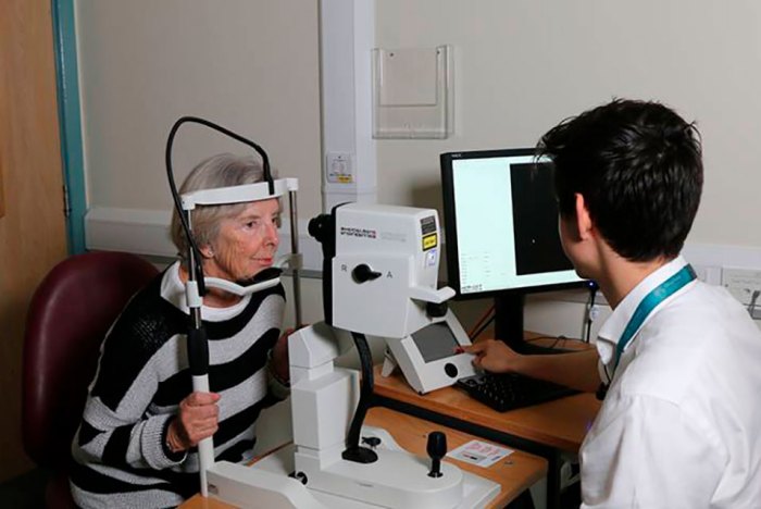
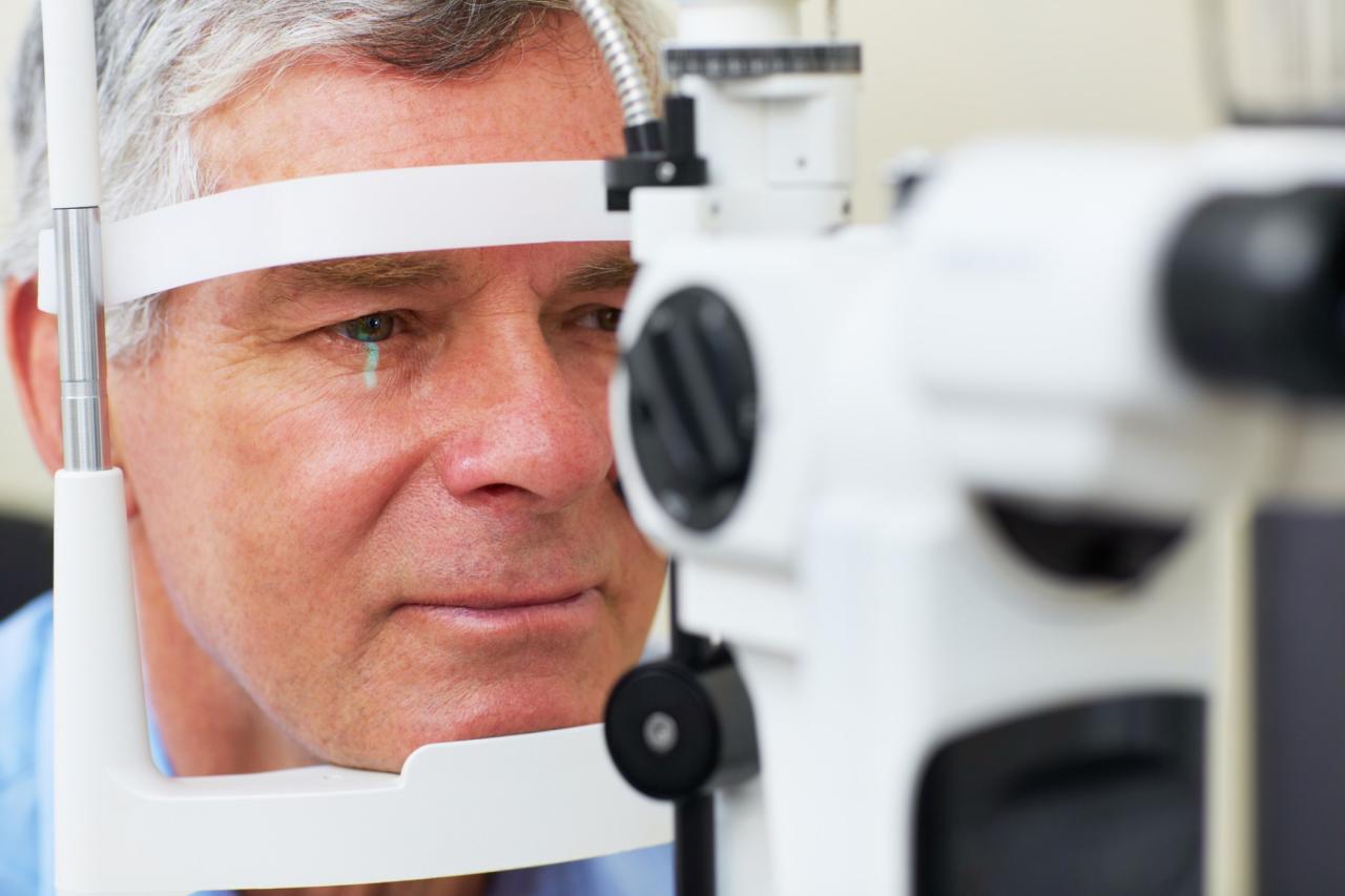
D eye scans, also known as optical coherence tomography (OCT), utilize a technique similar to ultrasound to create detailed images of the eye’s internal structures. A beam of light is projected into the eye, and the reflected light is measured to create a 3D image of the retina, optic nerve, and other structures.
Eye Structures and Parkinson’s Disease
Several eye structures and their associated changes have been linked to Parkinson’s disease. 3D eye scans can help identify these changes:
Retina
- Retinal Nerve Fiber Layer (RNFL) Thickness:Studies have shown that people with Parkinson’s disease may have thinner RNFL, which is a layer of nerve fibers that transmit visual information from the eye to the brain. This thinning may be related to the degeneration of dopamine-producing cells in the brain, a hallmark of Parkinson’s disease.
- Ganglion Cell Layer (GCL) Thickness:The GCL contains the nerve cells that process visual information. Studies suggest that GCL thinning might also be associated with Parkinson’s disease, potentially due to the loss of dopamine-producing cells.
Optic Nerve
- Optic Nerve Head (ONH) Size and Shape:Changes in the size and shape of the ONH, the point where the optic nerve connects to the eye, have been observed in individuals with Parkinson’s disease. This could be related to the disease’s impact on the central nervous system.
- Optic Nerve Head Blood Flow:Studies have shown that people with Parkinson’s disease may have reduced blood flow in the ONH, which could be a consequence of impaired blood vessel function or nerve damage.
Other Structures
- Macula:The macula is the central part of the retina responsible for sharp central vision. Some research suggests that changes in the macula, such as increased vascularity, might be associated with Parkinson’s disease.
The Science Behind the Connection: 3d Eye Scans Can Detect Signs Of Parkinsons
The potential of 3D eye scans to detect early signs of Parkinson’s disease is based on a complex interplay between the nervous system, eye health, and the progression of the disease. Understanding the intricate connections between these factors is crucial for comprehending how eye scans might offer valuable insights into Parkinson’s disease.
The Role of Dopamine
Dopamine, a neurotransmitter vital for movement control, plays a critical role in both eye function and Parkinson’s disease. In the context of eye health, dopamine is essential for regulating eye movements, focusing, and visual processing. Parkinson’s disease, however, is characterized by the progressive loss of dopamine-producing cells in the substantia nigra, a region of the brain responsible for motor control.
This dopamine deficiency leads to the characteristic motor symptoms of Parkinson’s disease, including tremors, rigidity, and slow movements.
Neurological Pathways Linking Eye Health and Parkinson’s Disease
The connection between eye health and Parkinson’s disease is rooted in the shared neurological pathways that govern both functions. The substantia nigra, the brain region affected in Parkinson’s disease, is connected to the brainstem, which controls eye movements. The loss of dopamine-producing cells in the substantia nigra can disrupt these connections, leading to changes in eye movements that may be detectable through 3D eye scans.
Find out further about the benefits of social problems blamed on video games france riots macron that can provide significant benefits.
Potential Mechanisms by Which Eye Scan Abnormalities Might Reflect Parkinson’s Disease Progression
Eye scan abnormalities may reflect Parkinson’s disease progression through various mechanisms:
- Changes in Eye Movements:The loss of dopamine in the substantia nigra can affect the smooth pursuit system, which allows the eyes to track moving objects. This can manifest as difficulties in maintaining a steady gaze, potentially detectable through 3D eye scans.
- Alterations in Pupillary Response:The pupil’s response to light can be affected in Parkinson’s disease. 3D eye scans can potentially measure changes in pupillary dilation and constriction, which might be indicative of dopamine depletion.
- Structural Changes in the Eye:Parkinson’s disease can cause subtle changes in the structure of the eye, such as the thickness of the cornea or the shape of the lens. 3D eye scans can provide detailed measurements of these structures, potentially revealing early signs of disease progression.
Research and Clinical Studies

The potential of 3D eye scans to detect Parkinson’s disease has been the subject of numerous research studies, with findings offering valuable insights into the diagnostic capabilities of this technology. These studies have explored the accuracy and reliability of 3D eye scans in identifying individuals with Parkinson’s disease, providing a foundation for understanding its potential role in clinical settings.
Accuracy and Reliability of 3D Eye Scans
The accuracy of 3D eye scans in diagnosing Parkinson’s disease is a critical aspect of research. Studies have explored the ability of these scans to distinguish between individuals with and without the disease, evaluating the sensitivity and specificity of the technology.
Sensitivity refers to the ability of a test to correctly identify individuals with the disease, while specificity refers to its ability to correctly identify individuals without the disease.
For example, a study published in the journal “Movement Disorders” in 2019 found that 3D eye scans had a sensitivity of 80% and a specificity of 75% in identifying individuals with Parkinson’s disease. This suggests that 3D eye scans can accurately detect the disease in a significant proportion of cases, while also minimizing the risk of misdiagnosis.
Potential Benefits of 3D Eye Scans
The use of 3D eye scans for Parkinson’s disease diagnosis offers several potential benefits.
- Early Detection: 3D eye scans have the potential to detect early signs of Parkinson’s disease, even before the onset of motor symptoms. This could enable earlier intervention and potentially slow down disease progression.
- Non-invasive and Accessible: 3D eye scans are a non-invasive and relatively accessible diagnostic tool, making them suitable for routine screening. This could facilitate early detection and diagnosis in a wider population.
- Objective Assessment: 3D eye scans provide objective measurements of eye movements, reducing the subjectivity often associated with clinical assessments. This can lead to more accurate and reliable diagnoses.
Limitations of 3D Eye Scans
Despite the promising potential of 3D eye scans, it’s important to acknowledge their limitations.
- Not a Definitive Diagnosis: 3D eye scans are not a definitive diagnosis for Parkinson’s disease. Further clinical evaluation and confirmation are still required.
- Limited Availability: 3D eye scanning technology is not widely available in all healthcare settings. This could limit its accessibility and potential for widespread adoption.
- Cost Considerations: The cost of 3D eye scans may pose a barrier to access, particularly in resource-limited settings.
Future Directions and Potential Applications

The potential of 3D eye scans for Parkinson’s disease goes beyond early detection. This technology holds promise for revolutionizing disease management and improving patient outcomes.
Monitoring Disease Progression and Treatment Response
D eye scans can be used to track changes in the eye over time, providing valuable insights into disease progression. This non-invasive method allows for longitudinal monitoring, enabling healthcare professionals to assess the effectiveness of treatments and adjust therapies as needed.
For instance, if a patient is undergoing medication therapy, regular 3D eye scans can help determine if the treatment is slowing or halting the progression of Parkinson’s disease. Similarly, if a patient undergoes deep brain stimulation surgery, 3D eye scans can monitor the impact of the surgery on disease progression and identify any potential complications.
Potential Challenges and Ethical Considerations
While 3D eye scans offer a promising tool for Parkinson’s disease management, several challenges and ethical considerations must be addressed for widespread adoption.
Cost and Accessibility
The cost of 3D eye scan technology can be a barrier to accessibility, particularly in resource-limited settings.
Standardization and Validation
Ensuring standardization of 3D eye scan protocols and validation of the technology across different populations is crucial for reliable and accurate results.
Privacy and Data Security
The collection and storage of sensitive patient data require robust privacy and data security measures to protect patient confidentiality.
Informed Consent and Patient Education
Patients must be fully informed about the risks, benefits, and limitations of 3D eye scans before consenting to the procedure. Clear and comprehensive education is essential to ensure informed decision-making.


